Novel microscope could be used for melanoma detection

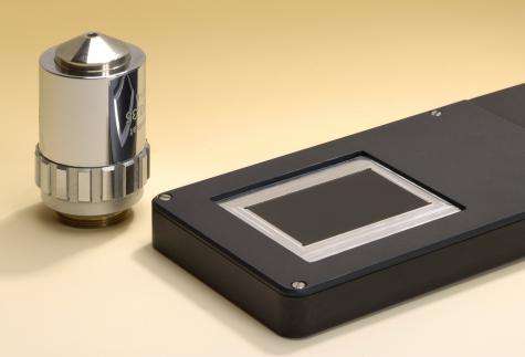
Suspicion of melanoma: In the future, doctors can pull out a new type of microscope to get to the bottom of suspicious changes in the skin. It provides a high-resolution image of skin areas of any size – and so quickly that you can hold it in your hand without blurring the resulting picture.
Are the dark spots on a patient’s skin malignant? In the future, doctors will be able to take a closer look at suspicious blemishes using a new microscope – with results in just a few fractions of a second. It examines to a resolution of five micrometers; it’s also flat and lightweight, and it records images so quickly that the results are not blurred even if the doctor is holding the microscope in his or her hand. For results with comparable resolution values, a conventional microscope would either be restricted to a tiny field forced to scan the surface: conventional equipment slowly sweeps the surface, point by point, recording countless images before combining them to create a complete picture. The drawback: it takes quite a while before the image is complete.
The new microscope designed by researchers at the Fraunhofer Institute for Applied Optics and Precision Engineering IOF in Jena, Germany, combines the best of both types of microscope: because it foregoes the grid, it needs to make just a single measurement, and that’s what makes it very fast. Still, it records across a broad imaging area. “Essentially, we can examine a field as large as we want,” remarks IOF group manager Dr. Frank Wippermann. “At five micrometers, the resolution is similar to that of a scanner.” There is also another benefit to the new system: With an optical length of just 5.3 millimeters, the microscope is extremely flat.
But how did researchers accomplish this feat? “Our ultrathin microscope consists of not just one but a multitude of tiny imaging channels, with lots of tiny lenses arrayed alongside one another. Each channel records a tiny segment of the object at the same size for a 1:1 image,” Wippermann explains. Each slice is roughly 300 x 300 µm² in size and fits seamlessly alongside the neighboring slice; a computer program then assembles these to generate the overall picture. The difference between this technology and a scanner microscope: all of the image slices are recorded simultaneously.
The imaging system consists of three glass plates with the tiny lenses applied to them, both on top and beneath. These three glass plates are then stacked on top of one another. Each channel also contains two achromatic lenses, so the light passes through a total of eight lenses. Several steps are involved in applying the lenses to glass substrates: first, the scientists coat a glass plate with photoresistant emulsion and expose this to UV light through a mask. The portions exposed to the light become hardened. If the plate is then placed in a special solution, all that remains on the surface are lots of tiny cylinders of photoresist; the rest of the coating dissolves away. Now, the researchers heat the glass plate: the cylinders melt down, leaving spherical lenses. Working from this master tool, the researchers then generate an inverse tool that they use as a die. A die like this can then be used to launch mass production of the lenses: simply take a glass substrate, apply liquid polymer, press the die down into it and expose the polymer layer to UV light. In a process similar to the dentist’s method of using UV light to harden fillings, here, too, the polymer hardens in the shape the die has printed into it. What remains are tiny lenses on the glass substrate. “Because we can mass-produce the lenses, they’re really pretty low-cost,” Wippermann adds.
Researchers have already produced a first prototype and will be showcasing it at the LASER World of PHOTONICS trade fair in Munich, from May 23-26. Boasting an image size of 36 x 24 mm2, this microscope can capture matchbox-sized objects in a single pass. It will be at least another one to two years before the device can go into series production, according to the researcher. The spectrum of applications is diverse: with this technology, even documents can be examined for authenticity.
Provided by Fraunhofer-Gesellschaft


