MSU lab manager shares microscopic images of dinosaur bones with growing audience

The thin slices of dinosaur bone that Ellen-Thérèse Lamm produces and the colorful photographs taken of these fossils under a Montana State University microscope continue to gain new audiences.
So far this year, the manager of the Gabriel Lab for Cellular and Molecular Paleontology at the Museum of the Rockies (MOR) has published research with MOR Curator of Paleontology and MSU's Regents' Professor Jack Horner in a French scientific journal, produced a 2012 MOR calendar, been featured in a trade magazine and been chosen as a finalist in an international photomicrography contest.
The microscopic structure of dinosaur frills at different stages of growth was described and analyzed in a paper Lamm co-authored with Horner. The paper was published in April in Comptes Rendus Palevol a French journal of paleontology and evolutionary sciences. Lamm produced the thin-section slides used in this research to capture the images for her and Horner's publication, which described the unique tissue growth strategies that Triceratops used to ultimately grow such a massive expanded frill.
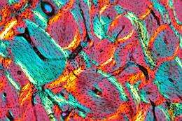
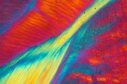
The photomicrographs were taken with a polarized light microscope, which enables scientists to manipulate light conditions to analyze the optical qualities of a sample, Lamm said. Polarized light passing through a thin-section of bone is split at different angles depending on the structure and organization of the crystal structures. It is then re-collected by an analyzer and delivered to the eye in a variety of colors and patterns.

"The images are not only beautiful and intriguing, but indicate different types of biological tissue, as well as show the orientation of fibers in the original bone," Lamm said.
This research further supported the MSU discovery that Triceratops and Torosaurus were actually the same type of dinosaur at different stages of growth, with Torosaurus being the mature adult stage of Triceratops, Lamm said. MSU graduate student John Scannella and Horner published that finding in July 2010 in the Journal of Vertebrate Paleontology. The discovery upended a long-standing belief that Triceratops and Torosaurus were completely different dinosaurs.
Some of Lamm's favorite images are also displayed in a 2012 fundraising calendar that she produced for the Gabriel Lab at the Museum of the Rockies. "Dinosaurs Under the Microscope - Paleohistology" includes images of dinosaur bone, modern animal specimens, as well as photos of MSU graduate students and Horner carrying out paleontological research. The calendar is now available online, in the Museum of the Rockies gift shop, the MSU Bookstore and downtown Bozeman at Country Bookshelf. To learn more, see www.lulu.com/spotlight/ET_Lamm .
Toward the end of the summer, the online trade journal called Delivering Performance ran a feature about the MOR paleohistology lab. The trade journal is produced by North American Composites, which donates the resin that Lamm uses to stabilize fossils before slicing them thin enough to examine under the microscope. An online version of the article can be viewed at www.nacomposites.com/deliverin … eid=18&page=interest .
Lamm was also a finalist in a photomicrography contest sponsored by Buehler Ltd, the world's premier manufacturer of scientific equipment and supplies used to analyze materials. The contest determines which photos taken through a microscope will be used in the company's 2012 calendar. Lamm's image showing the detailed microscopic structure of a Tyrannosaurus rex limb bone will be featured in August.
In addition, Lamm has just launched a website with extensive publication listings for Horner and colleagues, a Paleohistology Lab Blog, graduate student research, press stories, as well as a Google map showing the location of more than 50 fossil specimens from all over the world that were prepared at MSU. Since the Gabriel Lab is one of the only paleohistology labs in the world and is part of Horner's paleontology lab, researchers send their material here to be thin-sectioned by Lamm, who is one of the few people on the planet who does this kind of work. For more information, go to www.morhistologylab.org
Journal information: Journal of Vertebrate Paleontology
Provided by Montana State University


