New microfluidic chip can generate microbubbles to break open cells for biochemical analysis

Scientists have made many important discoveries in biology and medicine through studying the internal contents of cells. Some have isolated or identified nucleic acids or proteins with special functions, while others have unravelled the working and regulatory mechanisms underlying biochemical or pharmaceutical components within cells. Dave Ow and co-workers at the A*STAR Bioprocessing Technology Institute and Institute of High Performance Computing have now developed a novel method to expose the internal contents of cells for biochemical analysis.
Currently there is a wide range of methods to disintegrate or lyse cell membranes and to release the biomolecules contained within. However, most of these methods can cause denaturation of proteins or interfere with subsequent assaying. Ow and co-workers explored the possibility of using ultrasound in microfluidics to lyse cells. They applied short bursts of ultrasound with periods of rest to prevent the proteins from overheating as a result of dissipation of mechanical energy.
When the rapid changes of pressure generated with ultrasound are applied to a liquid, small bubbles are formed which oscillate in size and generate a cyclic shear stress. These rapidly oscillating bubbles generate a mini shockwave when they implode, which can be strong enough to cause the cell membrane to rupture. The researchers generated microbubbles in the meandering microfluidic channel by introducing a gas via a separate inlet to generate a gas–liquid interface and subsequently applying ultrasound to the system.
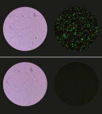
As a proof of principle, the researchers tested the performance of their microfluidic device on genetically engineered bacteria and yeast that express the green fluorescent protein. The researchers found that the bacteria are completely disintegrated after only 0.4 seconds of ultrasound exposure (see image). The concentration of DNA released from yeast cells reached a plateau after only one second exposure (which contained six bursts of ultrasound each of 0.154 seconds), indicating that most cells are successfully lysed. Importantly the temperature of the sample was shown not to rise above 3.3 °C. “The large surface to volume ratio of the microfluidic environment means that the small amount of heat that is generated rapidly diffuses away,” says Ow.
The researchers have proposed many ideas for applications. “In collaboration with another institute, we are developing a rapid and sensitive label-free optical method for on-chip detection of bioanalytes from lysed cells,” says Ow. “We also want to modify the device to break more difficult-to-lyse endospores, and to develop a rapid on-chip detection device to counter the threats of bioterrorism.”
More information: Research article in Lab on a chip
Provided by Agency for Science, Technology and Research (A*STAR)


