New technique lights up the creation of holograms

Researchers at the RIKEN Advanced Science Institute (Japan) have developed a unique way to create full-color holograms with the aid of surface plasmons.
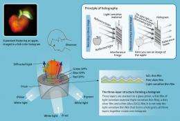
A red apple with green leaves—it seems real enough to pick up in one’s hand, but there is nothing there to actually touch. This is because the apple is a hologram, a three-dimensional image projected by light (Fig. 1). In April 2011, Chief Scientist Satoshi Kawata of RIKEN, the head of the Nanophotonics Laboratory at the Advanced Science Institute in Wako, along with his colleague Miyu Ozaki, a visiting scientist of RIKEN, succeeded in developing a novel holography principle distinct from conventional methods which makes it possible to reconstruct a full-color three-dimensional object. The key to their success resided in superposing a thin silver film on which surface plasmons—collective oscillations of free electrons within a metal—were excited. In recent years, research on surface plasmons has rapidly expanded against a background of remarkable advances in nanotechnology, helping establish the subject as a field of engineering. In addition to holograms, Kawata has also undertaken groundbreaking research on surface plasmon applications in metallic nano-lens technology.
Full-color holograms
Seven years ago during an interview with RIKEN News in February 2005, Kawata mentioned, “I want to do two things at RIKEN, one of which is to pioneer the new discipline of plasmonics.” Since then, he has authored several landmark publications in plasmonics, including one article which he coauthored with Miyu Ozaki, titled “Surface-Plasmon Holography with White-Light Illumination,” which appeared in Science (April 2011).
“In recent years, three-dimensional (3D) movies and television sets capable of 3D imaging have been gaining popularity, but the images of the objects we see are for the most part nothing more than pairs of planar images recognized as 3D objects by the brain due to the effect of lateral parallax,” Ozaki explains. “Meanwhile, holography has long been available as a classical technique for presenting 3D images. You may have seen a red or blue stereoscopic image floating in the air at an amusement park or science museum. However, it is difficult to create colored holograms because of the principle behind the technique. We have now succeeded in developing full-color holography using a new method. The red apple with green leaves shown in Fig. 1 is one example,” says Ozaki. Surface plasmons have enabled Kawata and Ozaki to produce holograms that are distinct from conventional ones.
A hologram is like a photograph that records the light waves scattered from an object, and reconstructs the object in three dimensions when the object is not actually present (Fig. 1). The image that we can see is the result of captured light that has collided with and been scattered by the object. “For recording, a laser beam is used, and is divided into two identical beams. The illumination beam lights up the object, and light reflected by the object is applied to a light-sensitive material, such as photo film. At the same time, another light source called a reference beam is superposed onto the photo-sensitive material,” explains Kawata. “The two beams interfere with each other and produce a pattern of dark and bright bands. This interference fringe contains information from the light scattered by the object, including information about the shape of the object, which is then recorded onto the photo-sensitive material or hologram. To reconstruct the object in three dimensions, ordinary light illuminates the pre-recorded hologram. The illumination is diffracted by the interference fringe pre-recorded in the hologram to regenerate the light wave scattered by the object during the recording. When we see the hologram under illumination, it looks as if the object is actually present.”
To obtain an exhaustive interference fringe, a laser that produces light waves with uniform crest-to-crest and trough-to-trough patterns must be used as the reference beam. The color of the diffracted light is influenced by the laser beam used, so the image reconstructed through a conventional hologram is the same color as the laser used. This is why most holograms are monochromatic. “Our newly-developed system employs laser beams for recording the hologram, but does not use any laser beams for the illumination beam that is required for image reconstruction. Instead, we use a beam of white light, which comprises a blend of many different wavelengths. Since the three primary colors of light—red, green and blue (RGB)—can be extracted from white light, stereoscopic images are reconstructed from holograms in the respective colors, and the three stereo images are superposed to obtain a combined full-color image,” Kawata says.
Extracting the three primary colors from white light using surface plasmon resonance
“The key to successfully extracting RGB separately from white light resides in superposing a thin silver film onto light-sensitive material. In doing so, surface plasmon polaritons (SPPs) are excited on the thin film,” says Ozaki.

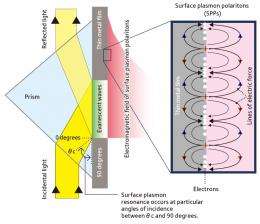
A metal contains a great number of free electrons that are oscillating together while simultaneously interacting with each other. The quantum of this collective oscillation of free electrons in a metal is called a plasmon. A plasmon is always accompanied by a photon or electromagnetic field. On metal surfaces, plasmons and photons propagate along the surface, and this combination is called surface plasmon polariton (SPP). Figure 2 illustrates how SPP works. When a beam of monochromatic light at a particular wavelength is applied to a thin metal film through a prism, it is totally reflected at angles of incidence between the critical angle (θ c) and 90 degrees. At the same time, dim light exists close to the boundary face. The resulting evanescent waves excite SPPs along the surface of the thin metal film. Usually, an SPP is not excited by the light incident on the metal film, except only at an angle for resonating SPPs. At a resonant angle, the energy of the incidental light transfers to the SPPs rather than to the reflected light. We use the evanescent light field associating with SPPs to illuminate a hologram.
Surface plasmon resonance is seen at given parameters of film thickness, material, incident angle of light, and wavelength of light. In the case of white light, which is a blend of many different wavelengths, the angle of incidence varies as a function of wavelength for RGB. “We make the best use of this mechanism. By changing the angle of incidence to a value that accommodates RGB, we can extract light beams in the three colors R, G and B separately from the same white light,” Ozaki says.
A powerful tool that will enhance communications technology
While the system diagram in Fig. 2 comprises only two components (a prism and a thin metal film), the hologram developed by Kawata and Ozaki (Fig. 1) has three layers. These layers comprise of a thin film of light- sensitive material, a thin silver film and a thin silica (SiO2) film stacked on a glass prism with undulations that correspond with the interference fringes for the three colors R, G and B on all three layers. A hologram is therefore not produced by the light-sensitive thin film alone, but by all the three layers as a whole. The thin silver film, which serves to facilitate color separation, is 55 nm thick, and the undulations matching the interference fringes are about 25 nm high. The total thickness of the three layers is several hundred nanometers.
“The three angles of incidence of white light (θ red, θ green, θ blue) have been adjusted in advance to allow surface plasmon resonance to occur for each of the R, G and B colors,” explains Ozaki. “A beam of white light applied in three directions, as shown in Fig. 1, excites the SPPs in each color, and the resulting electromagnetic fields are holographically diffracted to reconstruct a stereo image. Looking at it from above, the viewer can see a full-color stereo image.” The hologram created in this experiment measured 38 mm in length and 26 mm in width, with the subject being a fingertip-sized pendant featuring the image of an apple.

“This achievement will find applications in three-dimensional display devices in the future,” says Ozaki. “In the case of holograms generated using surface plasmons, white light alone serves as the incidental light, and beams can be applied from behind, so our system can be utilized in small devices such as smart phones.”
Ozaki notes that some problems remain to be solved before the system can be brought into practical use. “The system must be improved to allow larger objects to be imaged. In addition, the viewing angles for stereo images are now limited to about 25 degrees both vertically and laterally, so this range must be broadened. However, these engineering problems can be solved. Using our system, we also aim to create moving pictures. By using surface plasmons and holography in combination, numerous approaches are available, and we may later use a technical approach that is totally different from the technique we have just announced,” says Ozaki.
Metallic nano-lenses
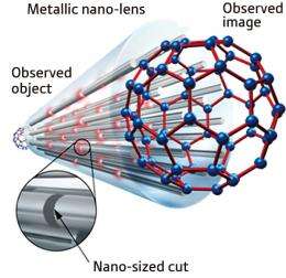
Prior to developing the full-color holography technique, in 2005 Kawata created a metallic nano- lens based on the principle of surface plasmons. The device is configured with bundles of nano-sized, thin metal wires and arranged in the form of a pin support. When an object is placed on top of the device, the light reflected by the object collides with the tips of the metal wires, producing surface plasmons. These plasmons carry information about the object and travel through the metal wires resulting in the reconstruction of an image on the opposite side. Due to the wires’ thinness, greater detail about the object can be transmitted, which produces a higher resolution of the reconstructed image. Theoretically, images can be transmitted at a resolution of 1 nm. However, the device created by Kawata and colleagues was only able to produce fixed-size images, and the colors of transferable images were limited to discrete wavelengths.
Later in 2008, Kawata published a new idea for a metallic nano-lens capable of providing enlarged images of objects at a resolution of several nanometers in Nature Photonics (Fig. 3). The new device is currently under fabrication. “The features of bundling many metal wires and transferring surface plasmons through metal wires are the same as those I announced in 2005,” says Kawata. Improvements include the capability of enlarging observed images to macroscopically visible sizes by arranging metal wires into a fan formation, and cutting the metal wires at some points to form nano-sized gaps. The gaps help to broaden the waveband, which can be covered. Information on the object is transferred through the wires as surface plasmons, and as light through the wire-to-wire gaps, and the plasmons are eventually released as light to image the object. In the case of gapless wires, the number of oscillations that permit surface plasmon resonance is subject to limitations, and the surface plasmons gradually lose oscillation energy. By providing gaps, the energy attenuation is prevented, and all wavelengths over the entire band of visible light are covered, so the object can be examined in full color. We have demonstrated this through theoretical calculations and computer simulations.”
Because the principle predicts that metallic nano-lenses will also work in water, further advances would enable examination of living cells on the nanoscale, offering expectations of applications in bioengineering and nanolithography in the future. “Plasmonics has a wide range of applicable fields, and some researchers are working on developing medical applications. For example, a cancer therapy may be feasible in which microparticles of silver-coated silica are delivered to the affected part of the body, and a beam of near-infrared rays is applied there to attack the cancer cells with plasmons. Because effective near-infrared rays are available in sunlight, sunbathing could possibly serve as a treatment,” says Kawata.
Watching single DNA bases using deep ultra-violet ray imaging
Kawata has been expanding his research area to encompass nanophotonics and plasmonics, and he is now focusing on deep ultra-violet ray plasmonics. “In the deep ultra-violet ray zone covering the wavelengths between 220 and 350 nm, gold and silver lose their metallic nature and no longer produce surface plasmons, while aluminum alone produces surface plasmons. If we realize deep ultra-violet ray imaging using an aluminum needle to narrow the surface plasmon resolution to 0.3 nm, we would be able to directly observe the base sequence of the DNA, which can be compared to the basic design of life. While many researchers study the terahertz band, which covers wavelengths of around 300 micrometers, I want to explore things that have not yet been explored by anyone else,” says Kawata.
Provided by RIKEN


