Researchers map signaling networks that control neuron function

In the first large-scale proteomics study of its kind, researchers at the University of California, San Diego School of Medicine have mapped thousands of neuronal proteins to discover how they connect into complex signaling networks that guide neuron function. Their research – using quantitative mass spectrometry, computational software and bioinformatics to match the proteins to their cellular functions – may lead to a better understanding of brain development, neurodegenerative diseases, and spinal cord regeneration.
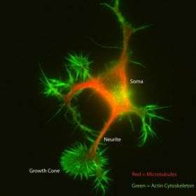
Led by Richard Klemke, Ph.D., professor of pathology at UCSD School of Medicine and the Moores UCSD Cancer Center, the research team designed a new technology enabling them to, for the first time, isolate and purify neurites – long membrane extensions from the neuron that give rise to axons or dendrites.
This technological breakthrough opens the door to understanding how neurites form and differentiate to regenerate neuronal connections and give rise to a functioning network. It also led to the discovery of how two key signaling molecules are regulated by a complex protein network that controls neurite outgrowth. Their study will be published the week of January 28 to February 1 in the on-line, early edition of the journal Proceedings of the National Academy of Science.
The formation of neurites, a process called neuritogenesis, is the first step in the differentiation of neurons, the basic information cells of the central nervous system.
“Understanding how neurites form is crucial, as these structures give rise to the specialized axons and dendrites which relay sensory input and enable us to see, hear, taste, reason and dream,” said Klemke.
Neurons regenerate by sending out one or several long, thin neurites that will ultimately differentiate into axons, which primarily receive signals, or dendrites, primarily involved in sending out signals. These long, branch-like protrusions have a specialized sensory structure called a growth cone that probes the extracellular environment to find its way and determine which direction the neurite should move in order to hook up with other neurites that will also differentiate into axons and dendrites.
The neural signaling network of dendrites and axons forms a huge information grid, which the UCSD team is studying in order to discover how neurons connect properly and regenerate to maintain proper wiring of the brain. Understanding the role that neuritogenesis plays in the regeneration of nerve connections damaged by diseases such as Alzheimer’s, Parkinson’s or other neurogenerative diseases is an important component of mapping the signaling network.
“Our primary goal is to identify unique proteins that cause the neurite to sprout and differentiate,” said Klemke. “We also want to understand the underlying signals that guide neurite formation and migration in response to directional cues.”
Klemke’s postdoctoral associates Olivier Pertz and Yingchun Wang identified a complex network of enriched proteins called GEFs and GAPs that control neuritogenesis by modulating signaling.
“This signaling provides external guidance cues to mechanical mechanisms inside the cell that make the neurite go forward, turn, or reverse direction,” Klemke said. “Understanding how the thousands of neurite proteins work in concert may someday help us guide neurites to the right place in the body to regenerate and reverse the impact of neural degenerative diseases or help facilitate spinal cord healing after injury.”
The researchers developed a unique microporous filter technology to separate the neurite from the cell body of the neuron, called the soma. The ability to slice millions of neurons into their soma and neurite components opened the door to using mass spectrometry, a tool able to identify the thousands of proteins that uniquely compose the two structures. Using information gleaned from published work, the researchers were then able to predict the function of most of the neurite proteins. This allowed them to construct a blueprint of how the thousands of proteins work together to facilitate neurite formation.
Source: University of California - San Diego


