An unusual collection : A brain tumor tissue bank

(PhysOrg.com) -- Five years ago, as she was walking into Caritas Holy Family Hospital and Medical Center in Methuen, Mass., Patricia Fey saw a priest she knew and cornered him. "I'm like 'Oh, Father Peter! And I sort of grabbed him by his arm," she recounts. "I said, 'What are you doing here? Father Peter! I could use a prayer right now. He asked me what was going on and I told him, "They found a brain tumor and I'm about to get set up for radiation. It's cancer."
“Father Peter put his hand on the top of my head, closed his eyes, and started saying a prayer,” Fey continues. But all she could think was, “Oh no! He’s blessing the wrong side!”
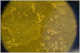
Today, a piece of the tumor the priest prayed over, along with many like it from other patients, is living in a laboratory at the Dana-Farber Cancer Institute, where researchers are studying the tissue samples, hoping to use them to tailor treatments for patients.
Harvard Medical School (HMS) neuro-oncologist Santosh Kesari and colleagues are beginning -- on an experimental basis -- to venture into the challenging and possibility-filled world of personalized medicine. “Studying (the tumor) outside of the body could be a personalized way to study the patient’s condition; a way to study the patient’s tumor as it evolves instead of studying a static sample that is already dead,” says Kesari’s colleague, Keith L. Ligon, assistant professor of pathology at Harvard Medical School and a consultant in neuropathology at Children's Hospital Boston.
Kesari, an assistant professor of neurology at HMS, and Ligon have established a tumor bank at Dana-Farber, which contains living tumors cultured from patients’ tumor cells.
“Part of the reason we started working on this tissue bank was just the obvious potential for using such cells for both research and potentially clinical care further down the line,” Ligon says.
“It’s like making a direct translation of science to the patient,” Kesari adds.
Kesari and Ligon first met six years ago when they were both postdoctoral fellows at the Dana-Farber Cancer Institute. Back then, they studied the development of glioblastoma brain tumors in mice. The work soon evolved into the analysis of this type of tumor in humans, assisted by new discoveries on ways to culture glioblastoma-type tumor cells. Researchers found a way to effectively isolate cells from these malignant masses, a discovery that led to the tissue bank project for Kesari and Ligon.
For Ligon, “the type of person who likes to collects things,” and who used to collect stamps and comic books when he was a boy, it was only natural. For Kesari, the project has recently taken the undertone of a personal crusade where his most important, though not the only, contender is time - a family member has been diagnosed with the disease.
For years, Kesari had remained a bench researcher, focused solely on doing basic science far removed from patient care. “But then you know patients are dying,” he says. “I realized that with the new technology, the genomics, the proteomics, there’s a possibility of bringing this science to the clinic. We see patients every day, and there are things we can learn from them. There is science that you can do just from the blood of patients undergoing treatment with specific drugs or the specimens that we get at surgery.”
Researchers have learned over the years that a brain tumor is not “one thing,” that every patient has a different “flavor,” as Kesari says, even in the way the illness presents itself.
Take Patricia Fey, for example.
In 2005, while participating of her daughter’s eighth-grade graduation, she began experiencing an intense headache. “I had been getting these headaches, lots of headaches,” she explains. “They didn’t go away. That day I couldn’t stand it. “It was like something pressing on my head.”
Fey was diagnosed with glioblastoma, one of the most common malignant brain tumors. Hers was a stage 4, the most lethal. Although the prognosis for a patient with glioblastoma usually is grim (12 months), it’s been five years since Fey’s diagnosis.
Ephraim Friedman, a physician from Beverly Farms, Mass., also became a patient five years ago when he was diagnosed with a brain tumor following a seizure that left him unconscious. Today, with slightly slurred speech and some issues with his short-term memory, Friedman is well enough to work on the sculptures he loves to create and says he is doing well.
These long-term survivals are great news for both Kesari and his patients. But Kesari and Ligon want to better understand the science behind these success stories. “They probably have a particular kind of glioblastoma that we don’t understand well enough,” Kesari says.
The problem, according to Kersari is "lumping" patients into one disease when “… the reality is, you look at the genetic profile of these tumors, they are very different from one another,” says the investigator. “Each person’s tumor is actually different even though it’s called the same. And each person responds differently to different drugs. So if we understood the connection between the genetic makeup of the tumor and the response to specific treatments that we have at hand, then we would be able to make a more rational treatment for the patients.”
Glioblastoma is also as nondiscriminatory as it is varied.
“This is one of the cancers that span all ages, from childhood to old age,” Kesari says, “That’s why it’s an important disease to understand, because it affects everyone.
“The common concept in the past was that a tumor was basically just a clone of itself,” Ligon says. “Yet in certain kinds of cancer, for example glioblastoma, when you look at the patient’s tumor, you notice cells don’t all look the same. It’s a heterogeneous mix when it starts.”
What researchers have found is that different cell types exist within an individual tumor. These cells may be highly genetically related, they may look different, or they may have different genetic makeup as well. It is this heterogeneity what makes “killing” the tumor so difficult. Drugs used in therapy can partially shrink the tumor by terminating a certain group of cells while other types of cells remain behind and replicate, thus maintaining the possibility that the tumor will propagate. One drug doesn’t target all cell types at once.
“The bottom line is that not every tumor cell is the same as its neighboring cell. That’s where the cancer stem cells come in,” Ligon says, referring to a controversial theory holding that not all tumor cell types are important for tumor growth.
“The question is what are the most important ones to kill within the tumor?” Ligon says.
The cancer stem cell theory, as it is known, is that there is a small population of “mother”cells with the potential to survive therapy. Much like the body’s normal stem cells, capable of generating the different tissues that comprise the body, these “mother” cells, or cancer stem cells, would reproduce infinitely turning into powerful, malignant “daughter” cells that can survive standard treatment that has not yet been designed to destroy their population.
The adult brain retains a certain number of stem cells over a person’s lifetime. These cells help regenerate the brain, and are possibly involved in memory and cognition, but some scientists think it is possible that, when lacking, they might also be causing degenerative diseases such as Alzheimer's, and Parkinson’s. “And the other flipside is,” Kesari says, “if they go wild, stem cells could also be involved in the formation of brain tumors. That’s something that’s not proven, though.”
And it’s a hard task because, if they exist, these sought-after cancer stem cells are thought to be extremely rare, making up approximately one in 100,000 brain cells. Currently, scientists have two preliminary ways to identify, isolate, and grow the suspected cancer stem cells.
The first involves dissociating the tumor in a culture dish. “The ones [cells] that continue to grow and make spheres, tumor neural spheres as we call them, are the ones that functionally are defined as cancer stem cells,” Ligon says.
The second involves inserting the cells inside the brains of mice with immunological systems that have been previously compromised to prevent them from rejecting the inserted human cells. “You put those cells in the mouse, and presumably the cells that grow out are the cancer stem cells,” says Ligon.
Kesari and Ligon have been growing their patients’ cells in the same media other stem cell scientists use to culture ‘traditional’ stem cells. So far, it only works about 50% of the time, and when it does, the medium keeps the suspected tumor stem cells alive and in the same state as they would be found inside the patient’s brain.
“Many people,” says Ligon, “are doing research to try to study (the cultured cells) based on their genetic characteristics or the markers on their cell surface with the idea of separating the different kind of cells and purifying them.”
Instead of giving the patient an ‘off the shelf’ treatment, a more specific ‘lumping’ of the disease would allow a physician scientist to take part of the tumor to a laboratory and test all available treatments on it in order to identify the one that would be most effective.
“This is like a test of sensitivity to different drugs…that’s the simple concept,” Kesari says.
A simple concept that the team of researchers is currently embarking upon by initiating a series of clinical trials with the potential to help, at least, a certain population of patients with glioblastomas made up of cells that exhibit a specific kind of surface marker.
Inside his office space, an excited Kesari points at his computer monitor showing a brain image of a patient’s glioblastoma tumor. “See this mass in the brain,” he says, “both sides of the brain should be the same but clearly there’s a big mass here causing a lot of problems to the patient. The patient had several surgeries, radiation, chemotherapy, and several other trial drugs. They failed every time.”
Having no other choice Kesari gave his patient a drug called Imaginibe, which has the effect of inhibiting the expression of a gene called pdgfr. “This drug surprisingly had dramatic effects. You can see the shrinkage of the tumor in one month, which is very unusual for a tumor because you know patients with these tumors die within a year, usually.”
The researchers were surprised to look at other patients who in the past had been treated with the same drug. “This drug was thought not to work,” Kesary says, “but in fact the few patients that responded had this gene amplified as well.”
What Ligon and Kesari found was not a miracle drug, but one possible way to predict whether a tumor would respond or not to a treatment. “… because when we looked at this further with some colleagues in pathology we found that in this patient’s tumor cells the expression of a gene that is inhibited by the drug was amplified.”
Kesari’s and Ligon’s inspiration for the upcoming clinical trial that will use the over-expression of the gene (pdgfr) as a marker of a specific kind of glioblastoma, began with their interaction with another of their patients. “We were treating this patient who responded well to the drug, Kesari says, “we tried of figure out why, and in the process found the biomarker.”
“Now, we expect not to be saying ‘oh, the drug failed,’” Kesari says. Knowing that only patients with specific markers would respond to the drug would allow Kesari and his team to be more efficient in their treatment. “…every patient [would] be screened [for the marker] so if only one in 10 guys has the marker, we’re going to enroll him on the trial and then we’ll find another 10 [like that] … you’d have people with the same marker, you’d treat them with these drugs, and probably 100 percent will respond now … It’s a concrete example of personalized medicine,” Kesari says.
These days Kesari’s project has turned more personal. When he started in the field six years ago, an uncle of his died of a glioblastoma. And now a very close aunt has been diagnosed with something that at least by “name” is the same illness. “She’s one of my favorite aunts. So you know, back then it was personal, but now this is very important for me to do,” he says. “So I’m doing everything I can to figure this out.”
Kesari is going “full steam ahead,” as he says. The culturing of the cells, the insertion in mice brains, the genetic testing - it will all get done. “I mean, she’ll get the standard treatment, but I’m going to try to do as much testing as I can to see whether there are any of the markers that can help us predict response,” he says.
“I’m trying to do it fast [despite] barriers that are always in the way. You know, there are a lot of barriers, including financial, institutional, etc. to try to hit the target,” says Kesari. “Everyone knows there’s a right thing to do but you need money to do it, you need infrastructure to do it, and you know, we're just doing it ourselves. I really have no choice. I mean, I can’t keep waiting for the infrastructure to be built to do it.”
And if he and his colleagues find something that really works? “The clinical trial will be one proof of concept. It the trial works, it will be big news. Then if we can find something for her [his aunt], we’ll certainly publish it and make it known for other patients. If we can find out what makes her tumor tick, what drug will work, that’s gonna be the basis for another trial to treat another subset of patients with that specific glioblastoma,” Kesari says. “That’s the only way to do it. You have to look at each patient, each tumor, because each one is different. It’s not just one disease. We’ve learned that from a lot of other cancers. You just need to do it in a brute force fashion, I think.”
Provided by Harvard University (news : web)


