Visualizing brain processes with new techniques

(PhysOrg.com) -- The brain's magic is worked by neural circuits, where information is transmitted from one nerve cell to the next. In the heat of the summer, for example, our ability to relish an ice cream is due to these circuits: they underlie our perception of its refreshing coolness, enticing color and smooth taste.
Unfortunately, however clear our perceptions may be as we enjoy that ice cream, disentangling the neural circuitry is anything but straightforward. As FMI neurobiologist Botond Roska admits, "We know a fair amount about the larger-scale anatomy of the brain, and we also understand some of the functions of the nerve cells themselves. But we know practically nothing about the detailed wiring, the connections, between cells."
Two tools developed and refined by Botond Roska's group at the Friedrich Miescher Institute now make it possible to identify and characterize neural circuits cell by cell. These new techniques are described in contributions to the prestigious journals Nature Neuroscience and Nature Methods and - given their major potential for use in neurobiology - they have been received enthusiastically by the research community.
Active nerve cells in all the colors of the rainbow
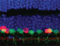
In a first set of experiments, Roska and collaborators from the University of Szeged (Hungary) demonstrate that it is possible to molecularly stain nerve cells in a circuit. This is achieved with the aid of a virus that "jumps" from neuron to neuron so long as the cells are interconnected synaptically. In the cells, the viruses produce two proteins - yellow for an active circuit and blue for inactive - that fluoresce more or less strongly according to the intensity of activation. The researchers were thus able to follow the transmission of information from neuron to neuron under the microscope.
They then went one step further: because a circuit frequently handles different types of information, and the individual components are used by several distinct circuits, the researchers used a whole palette of different colors for labeling. This allowed them to show how individual circuits drift apart and come together again and in which brain regions they end up. With this tool, Roska and his team can render circuits visible to an extent that was previously inconceivable.
Finally tackling persistent questions
The challenge lies not only in the sheer number of neurons and their interconnections. Different functions are taken on by different types of neuron For example, around 50-60 different neuronal cell types are found in the retina. Some are activated by specific colors, others only by the edges of objects, and still others respond to stimuli moving in specific directions. All these cells are interconnected in a variety of circuits that process the visual input. In the latest issue of Nature Neuroscience, Roska and his colleague Nathaniel Heintz of Rockefeller University, New York describe an elaborate study in which they identified mouse lines where green fluorescent protein is selectively expressed in specific cell types in the retina. The cell types can thus be precisely identified under the microscope. By combining this technique with the other new tool, it was possible to identify circuits activated in response to a particular visual impression.
Roska comments: "With these new techniques we can start to tackle questions that so far were beyond our means. We can now not only understand what happens in the brain after a particular stimulus, but for the first time we can also ask how neural circuits compute particular brain functions." Only when the - often densely packed - circuits of the nervous system are understood will it be possible to link complex human perceptions to physical processes in the brain.
More information: Boldogkoi Z et al. (2009) Genetically timed, activity-sensor and rainbow transsynaptic viral tools. Nature Methods 6:127-130
Provided by Friedrich Miescher Institute for Biomedical Research


