July 5, 2010 report
Latest imaging techniques look inside a python (w/ Video)
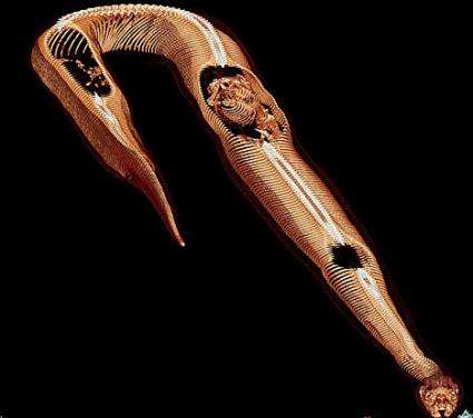
(PhysOrg.com) -- Researchers from Denmark have used CT scans and MRIs to see inside a python after it has swallowed a rat whole.
The scientists from the Department of Zoophysiology and MR Research Center at Aarhus University in Denmark, Henrik Lauridsen and Kasper Hansen, carried out computer tomography (CT) scans and magnetic resonance imaging (MRI) of a 5 kg Burmese python starting an hour after it had swallowed a whole rat. Contrast agents were used to highlight particular organs and show them in different colors. The snake was anesthetized before the scans.
The Burmese python (Python molurus bivittatus) is the largest sub-species of the Indian python, and is native to Southern and Southeast Asia. It fasts for months and then eats a large meal that can take several days to be fully digested. The meal can represent up to half of the snake’s own body weight. Wild pythons can reach over five meters in length and large snakes can consume animals as big as a deer.
The MRI study revealed how the python’s internal organs changed during digestion of the rat, which took 132 hours in total. As the body of the rat gradually disintegrated the snake’s gall bladder reduced in size, while the intestines expanded and its heart grew 25 percent larger.

Mr Lauridsen said the heart’s increase in size was probably related to the energy required for the digestion. Since the snake is a “sit and wait” predator that can go for long periods without eating, when it does eat the intestinal system must be restarted quickly.
The study, which was presented at the annual meeting of the Society for Experimental Biology in Prague, Czech Republic, enabled the scientists to see inside the snake without dissecting it, and thus avoided the sometimes subjective or misleading interpretations obtained from dissection. Dr Hansen also pointed out that dissection can induce changes, such as the collapse of lungs as a turtle shell is opened.
Since the study used a live snake, it could be studied “over and over again.” The images obtained should prove invaluable for future studies of anatomy of animals.
The study has already produced internal images of other species, including alligators, turtles, frogs, bearded dragons, and swamp eels.
© 2010 PhysOrg.com


